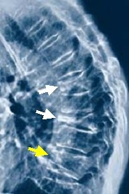
X-ray

X-ray examinations for osteoporosis or suspected osteoporosis are usually limited (in addition to the bone density measurement and of course if a bone fracture is suspected) to the spine, in particular the thoracic (thoracic spine) and lumbar spine (lumbar spine) and in particular to exclude or detect a vertebral fracture. In the case of acute, unclear back pain or a significant loss of size of more than 4 cm, x-rays of the spine should always be made in 2 planes.
It is not uncommon for a vertebral fracture to cause pain and is therefore overlooked. Since the risk of further vertebral fractures increases massively after one and with each subsequent vertebral collapse, the X-ray examination of the spine is of great importance!
* Image: White arrows: so-called osteoporotic compression fractures Yellow arrow: osteoporotic wedge vertebra (collapse at the front edge)
